Prerequisites
The Biological Bottom Line
Tissue repair follows an invariant chronological program: Hemostasis, Inflammation (Demo Day), Proliferation (Framing), and Remodeling (Curing).
Skipping inflammation with ice and high-dose NSAIDs leaves necrotic tissue debris in place, preventing cellular signals from building structural integrity.
Muscle heals in weeks because it is drenched in capillaries. Tendons and ligaments take months because they are largely avascular bradytrophic tissues.
The body first lays down fragile, disorganized Type III collagen as quick structural glue before slowly remodeling it into high-tensile Type I parallel cables.
If careful rest were the same thing as repair, a tendon would not still grab in month nine.
Yet people experience this constantly: you hurt an Achilles or patellar tendon, stop running, avoid stairs, and wait. Nine months later, the tendon catches on the first morning squat with the exact same ache. The common assumption is that the tissue is permanently broken or that aging closed the recovery window.
The calendar is the wrong object. Days flipping on a wall do not repair tissue. Healing is an active sequence of overlapping construction stations. If a handoff between stations stalls, the ticket sits untouched. You can wait three seasons in complete stillness, but if an earlier station never finished, the tendon remains unfinished.
The wall clock does not advance the ticket
Rest has a purpose. Unloading an injured structure stops picking the scab and prevents fresh tearing while the body organizes its response.
Rest is also incomplete. Tendons and ligaments do not expire into health from ice and waiting. Think of an order ticket pinned above the line in a commercial kitchen: prep, grill, garnish, pass. The ticket does not advance because the clock moved. It advances only when one station finishes its work and hands the plate forward.
Garnish a raw steak to save time and you ruin the meal. Skip prep and the grill has nothing to cook.
Tissue repair follows this logic. In Guide 1, we established that peptides are biological passwords, not cargo. A peptide cannot skip three stations because you are impatient. It is a signal that plugs into whichever station is already running. If that station is inactive, or the previous station never completed its handoff, the signal has nowhere to go.
The 4-Phase Tissue Repair Schedule
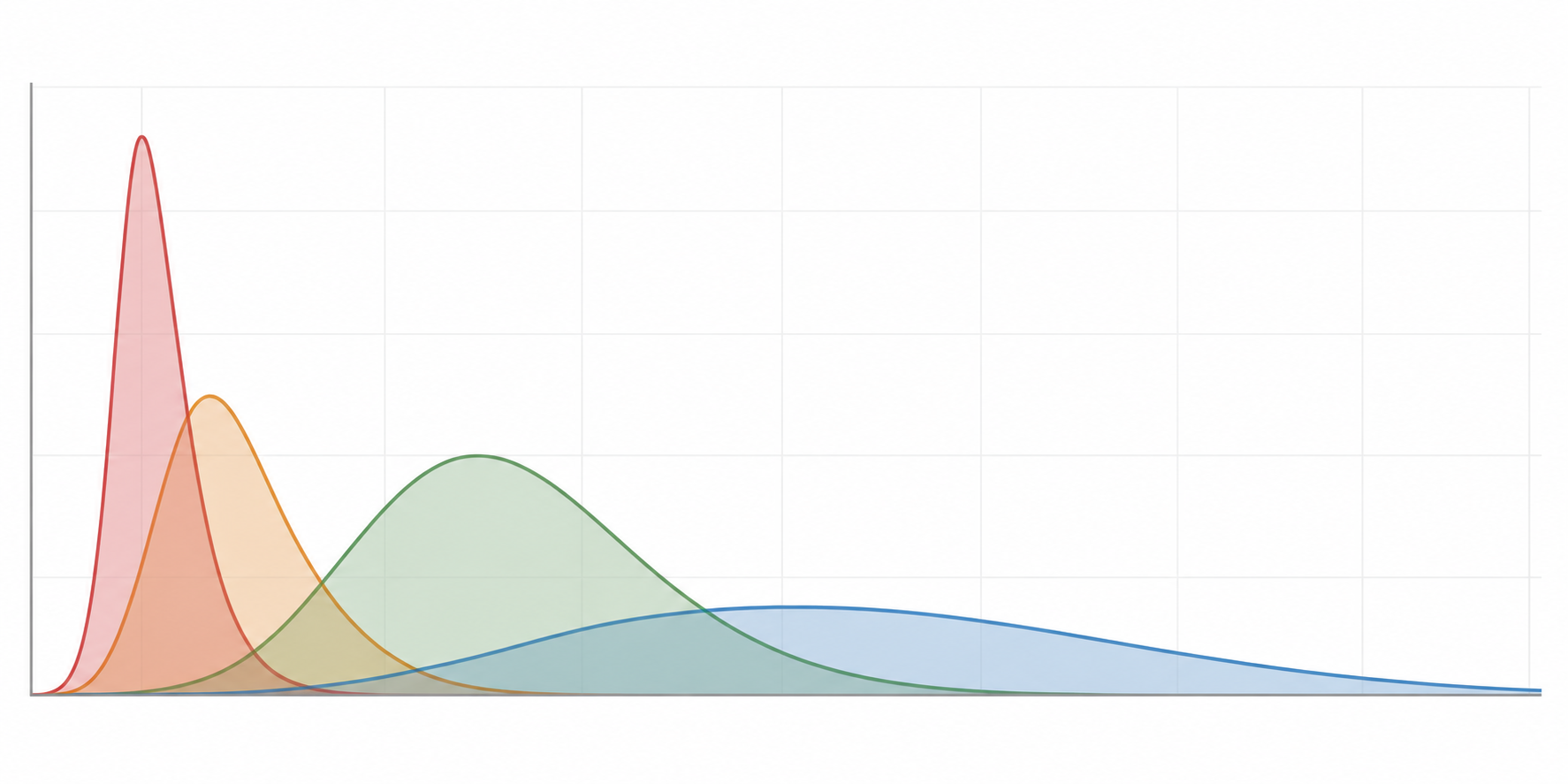
Phase 01: Hemostasis (Minutes to Hours)
Vascular constriction followed by platelet aggregation and fibrin clot formation. Creates a temporary matrix scaffold and releases primary chemoattractants (PDGF, TGF-beta) to summon the immune debridement team.
Four stations, overlapping on purpose
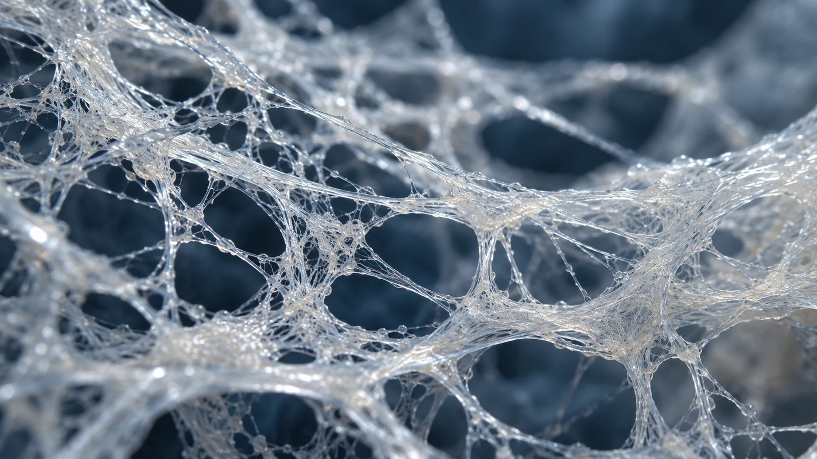
Living tissue does not punch a time clock between phases. The stations overlap, with the tail of one powering the start of the next.
The body runs through four stations in order:
Hemostasis stops the leak. When blood vessels tear, smooth muscle constricts to limit blood loss. Platelets gather at the breach, stick together, and weave a temporary mesh out of fibrin, a threadlike clotting protein. This plug seals the gap, while platelets release chemical call signals summoning the cleanup crew.
Inflammation is demolition and sanitation. White blood cells follow chemical signals to digest dead tissue, clear broken fibers, and establish chemical gradients for the rebuild. Many people suppress inflammation with ice and high-dose painkillers because it feels hot and swollen. Blunting symptoms does not skip biology. Eliminating cleanup forces the body to build on uncleared rubble. Because this phase is widely misunderstood, inflammation gets its own guide in Guide 3.
Proliferation builds the temporary frame. Once debris clears, fibroblasts (repair cells) migrate into the wound. To feed them, the body initiates angiogenesis, sprouting new blood vessels from existing capillaries. Fibroblasts manufacture collagen, the structural protein providing tensile strength. The body first lays down Type III collagen: a thin, pliable protein mesh. Type III collagen acts like biological duct tape, bridging the gap quickly but lacking the tensile stiffness to handle heavy loads.
Remodeling replaces the temporary frame with permanent architecture. The body breaks down fragile Type III collagen and replaces it with Type I collagen: thick, stiff, bundled protein cords built to withstand massive tensile force. During remodeling, these fibers align parallel to lines of stress and cross-link for durability.
In plain words:
- Hemostasis: seal the leak and post the first chemical call.
- Inflammation: clear debris and establish the repair zone.
- Proliferation: sprout temporary blood pipes and weave a fast, fragile mesh.
- Remodeling: replace the fragile mesh with aligned, high-strength cable.
Why tendons lag behind skin
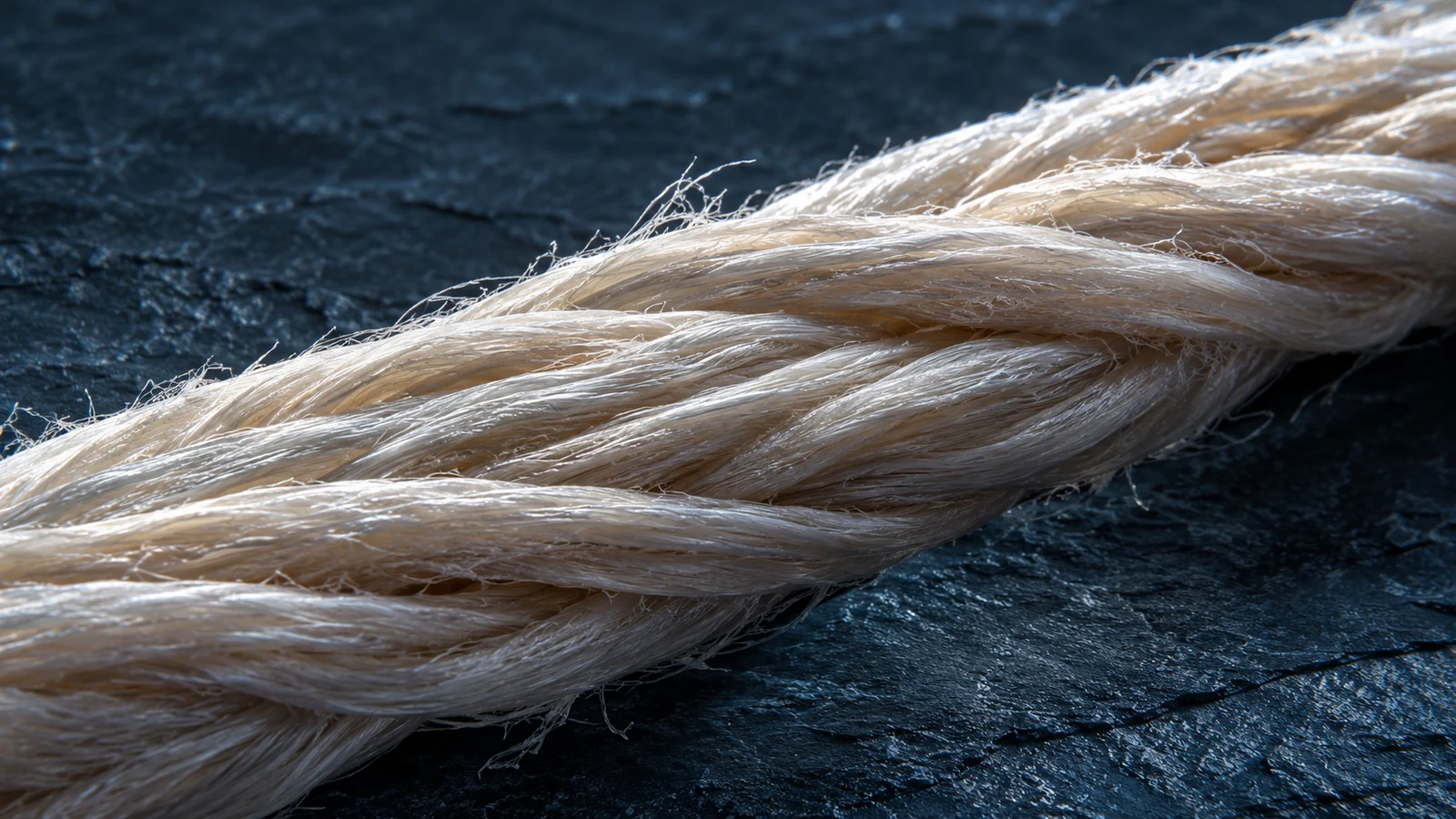
A finger cut heals in days. An injured tendon takes months.
Skin is packed with blood vessels and repair cells, maintaining constant access to oxygen and nutrients to build a simple, watertight barrier.
Tendon performs brutal mechanical work. It is a high-tension cable transmitting force from muscle to bone. To maintain density, tendon is naturally avascular, meaning it lacks a direct blood supply. It is also bradytrophic, meaning it has very low metabolic turnover and minimal oxygen consumption.
Because tendons rely on sparse vascular watershed zones, delivering raw materials is slow. Angiogenesis takes time to push new capillaries into dense fibrous tissue. Without pipes, you have no lumber; without lumber, proliferation and remodeling stall. Muscle heals in weeks because it is drenched in capillary blood. Tendon and ligament take months, and poorly supplied cartilage can take years, because blood access governs repair speed.
Where a ticket stalls
When an injury becomes chronic, the tissue did not lose its blueprint. The ticket stalled at a specific station.
A ticket usually stalls for three reasons:
Access. Hypovascular tissue cannot import enough immune cells for demolition or fibroblasts for reconstruction. The sequence halts because the supply line is dry.
Metabolism. High blood sugar and systemic inflammation alter collagen chemistry. Excess glucose binds to structural proteins, making collagen brittle. Systemic inflammatory noise also prevents immune cells from cleanly shifting from demolition to construction. A blood-sugar problem is not a side quest sitting next to the tendon. It directly degrades the material used to rebuild.
Load errors. Remodeling cannot occur without mechanical force. Total rest starves remodeling of its alignment cue, leaving collagen in a disorganized patch. Heavy loading applied too early during proliferation tears the fragile Type III mesh, resetting the ticket back to hemostasis.
Scar tissue is a speed-over-perfection compromise: disorganized collagen thrown down when the body needed immediate closure. Remodeling can replace much of that patch with aligned, high-tensile cords, but only if blood supply, chemical signals, and mechanical tension show up together. Without remodeling, the messy scar remains permanent.
Collagen Type III vs. Type I Maturation
Type III Collagen (Reticular Glue)
Secreted rapidly by early fibroblasts during the proliferative phase (Days 3 to 21). Thin, randomly oriented, highly pliable fibers. Provides fast mechanical continuity but lacks high-load tensile stiffness.
Type I Collagen (Parallel Cables)
Replaces Type III during the remodeling phase (Months 2 to 12). Thick, bundled triple-helix cables aligned strictly along the primary vectors of mechanical tensile stress through progressive loading.
Skeletal Muscle
2 to 4 Weeks. Rich capillary beds provide constant oxygen, amino acids, and immune access. Heals rapidly with minimal scar tissue under proper loading.
Tendon
12 to 24 Weeks. Bradytrophic tissue. Blood supply arrives via sparse paratenon vessels. Requires mechanical tension to drive fluid shear and signaling.
Ligament
24 to 52 Weeks. Dense fibrous bands with even fewer fibroblasts and blood vessels than tendons. Healing takes up to a full year to regain tensile stiffness.
Articular Cartilage
Years / Irreversible. Zero direct blood vessels or lymphatic channels. Chondrocytes receive nutrients purely via synovial fluid diffusion during joint compression.
Mechanotransduction: load is an instruction
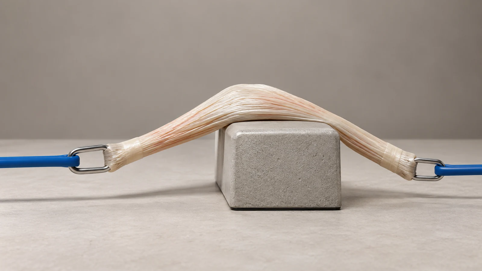
The body does not align collagen cables by checking a calendar. It aligns them through mechanotransduction.
Mechanotransduction is the biological process by which living cells sense physical tension and convert that mechanical force into chemical instructions. When you place controlled tension across a healing tendon, specialized tendon cells called tenocytes stretch. That stretch signals cells to synthesize Type I collagen and lay fibers down parallel to the line of pull.
Load is not just exercise; load is the architectural instruction that tells the construction crew which way the cable must face.
A chemical signal that says "make collagen" without a physical load that says "make it face this way" creates useless filler instead of a functional cable. Meanwhile, cells cannot build protein out of thin air. Dietary protein supplies the amino acids, specifically glycine and proline, that form the collagen strand. The biological signal, the mechanical load, and the amino acid substrate are all required. None is optional.
A peptide cannot skip a station
Peptides are signaling molecules, not regenerative erasers. They attach to cell receptors and request an action, such as accelerating angiogenesis or recruiting fibroblasts. Those requests only work if the target station is currently operational.
If an injury is trapped in unresolved inflammation, signaling for cell migration builds on debris. If proliferation has woven a fragile Type III mesh, but you avoid progressive load, no peptide can force those fibers to align into Type I cables. If your diet lacks amino acids, the instruction to build arrives without materials.
Key idea
The limits of a peptide are straightforward: wrong phase, broken handoff, absent load, or lack of substrate. A peptide is not Type I collagen, and it is not a loaded squat. It cannot replace the sequence.
What to ask when the calendar is lying
When an injury drags on, stop asking how long it has been. Ask which station looks stalled by looking at the physical behavior of the tissue:
- If the site is persistently warm, swollen, red, and irritable, inflammation has not finished demolition or resolved its chemical alert.
- If the site is quiet, cold, stiff, and weak, access and proliferation never took off. Blood supply and cell migration failed to establish a bridge.
- If the tissue feels stable at rest but catches or aches on the first loaded movement of the day, proliferation laid down the temporary mesh, but remodeling never aligned the fibers into a cable.
Do not use these distinctions to self-diagnose cellular phases or build a shopping list of vials. Bring them to a sports medicine clinician or physical therapist. Ask what objective signs show that cleanup finished, that blood supply reached the zone, and that the tissue is ready for progressive loading to drive remodeling.
Time was never the cook
The kitchen ticket was a translation, not the biology. Put it down.
Time does not knit tissue. Four overlapping stations (hemostasis, inflammation, proliferation, and remodeling) must each complete their work and hand the plate forward. Type III collagen is emergency tape. Type I collagen is finished cable. Mechanotransduction tells collagen which way to face. Dietary amino acids provide the raw lumber. A peptide is a signal, not a substitute for the cascade.
If rest, ice, and waiting could run the ticket on their own, month nine would not still grab. Time was never the cook.
Time was never the cook.
Educational content only. It is not individual medical advice.